Histological staining is used to highlight important features of the tissue as well as to differentiate structural elements of the tissue by their color and/or staining intensity. They are classified into 2 categories: Routine and Special Stains.

Histological staining

Routine Stain – Hematoxylin and Eosin (H&E)

At the Histology Core we routinely stain slides with the Hematoxylin and Eosin (H&E) stain. It provides a comprehensive picture of the microanatomy of a tissue and is frequently used by pathologists and researchers as an initial assessment.

How does H&E staining work?

It consists of 2 dyes; Hematoxylin and Eosin, which will stain the nucleus and the cytoplasm, respectively, in two different colors. Hematoxylin is a basic dye that stains the acidic components of the tissue (e.g., nucleus, ribosome, and rough endoplasmic reticulum) a purplish-blue color.  Eosin is an acidic dye that stains the basic tissue components (e.g., cytoplasm, cell walls, extracellular fibers) a reddish or bright pink color.

H&E Stain (left) vs Prussian Blue iron stain (right) in mouse spleen
PAS Stain (left) vs Silver Stain (right) showing basement membrane in the glomeruli
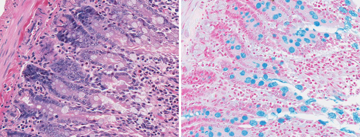
H&E Stain (left) vs Alcian Blue Stain (right) showing goblet cells in the rat intestine

Special stains

Special stains are usually performed after H&E staining. It refers to a wide variety of alternate staining techniques and procedures used to provide more in-depth information on specific structure(s) in a sample. Rather than specific antibody binding, special staining techniques are based on simple chemical reactions such as acid-base chemistry and oxidation-reduction reactions.

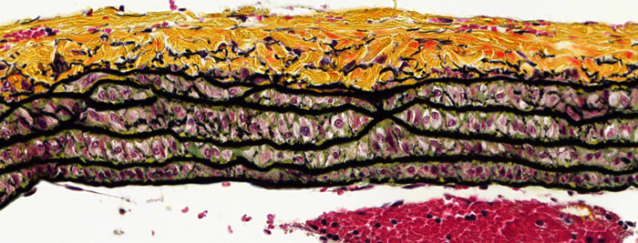
Movat Pentachrome Staining in rat lung
Movat pentachrome staining in rat lung

The Histology Core Facility routinely offers the following Special Stains:

If you would like to have a stain that is not listed, please email pathlab@uottawa.ca to enquire.

Carbohydrates

  • Periodic Acid-Schiff (PAS)
  • Periodic Acid-Schiff (PAS) diastase (Glycogen)
  • Alcian Blue (pH 0.4, 1.0, 2.5)

Connective tissue

  • Masson’s Trichrome (collagen, fibrotic tissue, cartilage)
  • Movat’s Pentachrome (collagen, elastic fibers, mucins, muscle, cartilage)
  • Verhoeff-Van Gieson (Elastic)
  • Picrosirius Red (Collagen)
  • Safranin O (Cartilage)
  • Toluidine Blue (Cartilage, Mast cells)
  • HPS (Hematoxylin Phloxine Stain: Muscle, Collagen)

Lipids

  • Oil Red O – Frozen tissue only

Microorganisms

  • Gram stain, Brown and Brent
  • Modified GMS Silver Stain (Fungi)
  • Giemsa Jenner (Parasites)

Hematology

  • Wright Giemsa (Blood Smear)

Minerals and pigments

  • Perl’s Prussian Blue (Iron )
  • Von Kossa (Calcium)
  • Alizarin Red (Calcium)
  • Congo Red (Amyloid)

Nervous system

  • Cresyl Violet (Neurons)
  • Luxol Fast Blue (LFB), LFB –PAS  (White and Grey matter)

Other stains (contact the Core if you require other stains)

  • Churukian Silver Stain (Alpha cells in pancreas, neuroendocrine cells)
  • Gomori’s Aldeyhyde Fuchsin (Beta cells in pancreas)
  • Jones Methenamine Silver ( glomerular capsule)